The eye (from the Latin word oculus, eye) is the organ of vision. By capturing light, it allows us to interact with the environment. The human eye can distinguish shapes and colors.
eye anatomy
The adult eye, or eyeball, is a sphere about 2.5 cm in diameter. It is protected in front by the eyelids, each of which bears eyelashes. They are lined by a membrane, the conjunctiva, which secretes a lubricating mucus to prevent the eye from drying out. The lacrimal gland, located above the eye, continuously releases tears (about 1mL/day/eye) helping to clean and protect the eye.
The bulb of the eye
The eye itself is called the bulb of the eye. It is a hollow sphere filled with liquids whose wall is made up of three layers (envelopes) superimposed. The transparent lens is the « lens » of the eye, it focuses light on the retina. It is protected by a liquid called aqueous humor.
The outer envelope of the eye is a fibrous tunic. It is a protective membrane which is composed of two elements: the sclera and the cornea. The sclera is the “white of the eye”. The cornea is transparent, it is the most exposed part of the eye. She very often suffers injuries but her ability to regenerate and heal is extraordinary. The cornea can also be transplanted without risk of rejection because it does not contain any blood vessels.
The intermediate envelope is a vascular tunic which is made up of three elements:
- The choroid, behind the eye, is a membrane rich in blood vessels which nourishes the three ocular tunics. It contains a dark brown pigment that absorbs light preventing its reflection inside the eye.
- The ciliary body is located in front of the eye, in continuity with the choroid. Composed of ciliary muscles, it acts on the shape of the lens by adapting its curvature to the distance from objects.
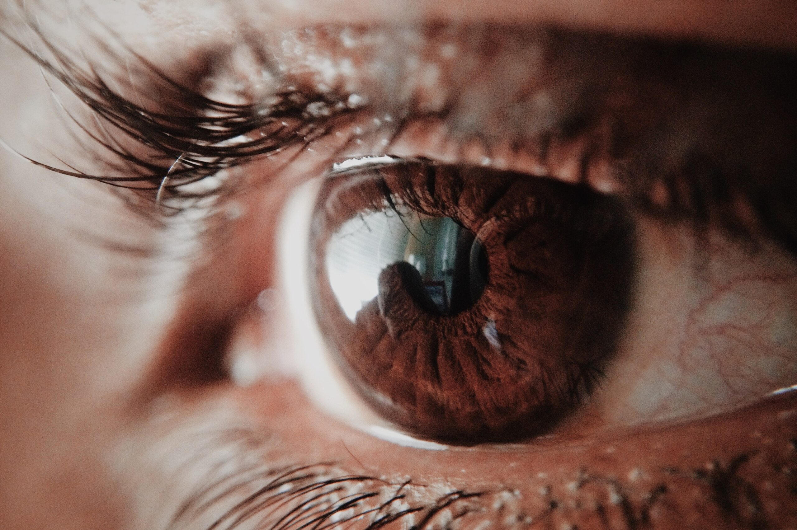
- The iris is located between the cornea and the lens. It contains pigment cells that determine the color of the eyes. In the center is the pupil, a round hole that lets light into the eye.
The inner coat of the eye is the retina. It extends from back to front, to the ciliary body. Half a millimeter thick, it is composed on its outer side of a pigmentary layer, glued to the choroid, which absorbs excess light thanks to melanin granules. Its inner side contains millions of visual cells: rods and cones. In its center, there is a small area called the macula : this is where visual acuity is maximum.
Visual cells and light in the eye
Visual cells
Visual cells are called photoreceptor neurons because they react to light.
The rods (120 million in each retina) are largely located at the periphery of the retina. They are responsible for peripheral vision and make it possible to distinguish shades of gray in the dark.
The cones (6 million) are mainly located in the center of the retina. They are involved in the vision of colors and details in bright light.
There are three types, each of which detects different wavelengths of visible light:
- The cones of the first type react mainly to blue light
- The second type cones in green light
- Third type cones react to red light
When excited by light, photoreceptors translate light energy into an electrical signal. These visual messages leave the retina via the optic nerve to reach the brain where they will be interpreted to produce visual sensations.
Light path in the eye
When light enters the eye, it successively passes through the cornea, the aqueous humor, the lens and the vitreous body, then reaches the retina in order to stimulate the photoreceptors. The image projected on the retina is inverted and smaller than the observed object.
Pathology and diseases of the eyes
The main global cause of visual impairment is cataracts with nearly 47.9% cases. Other major causes of visual impairment include glaucoma (12.3%), age-related macular degeneration (8.7%), diabetic retinopathy (4.8%) and trachoma (3.6%). )³.
Retina
Total achromatopsia : insufficiency of the three types of cones which results in a total absence of color vision. The vision is in shades of grey.
Daltonism : insufficiency of one of the types of cones which prevents distinguishing certain colors, more frequently red or green.
Age-related macular degeneration (AMD) : degeneration of the cells of the macula which results in loss of central vision (peripheral vision is not affected).
Retinal detachment : due to a small pocket of fluid located under the retina, it mainly follows a tear in the retina which allows intraocular fluid to infiltrate and lift it. The affected area no longer functions.
Diabetic retinopathy : damage to the retina resulting from retinal vascular disorders that can occur in people who have had diabetes mellitus for several years.
Vision disorders
Myopia : clear near vision but blurred distance vision.
Hyperopia : blurred near vision.
Presbyopia : decrease in near visual acuity.
Astigmatism : blurred near and far vision.
Lens
Cataract : opacification of the lens hindering the passage of light, which blurs near vision. First cause of blindness in the world.
Eyelids
Stye : infection of the base of the eyelid after infection by a bacterium.
Trachoma: infectious disease caused by the bacterium Chlamydia trachomatis , initially affecting the eyelid and which can cause blindness after repeated infections. Occurrence of disease in crowded places with limited access to water and health care. Spreads rapidly through contact with the hands or clothing of an infected person.
Eye disease
Glaucoma : damage to the optic nerve resulting in loss of visual field.
Dry eye syndrome : due to a decrease in the secretion of tears or an imbalance in its composition.
In the child
Visual impairment : accounts for 1% of births. Babies don’t stare at their toys and don’t respond to smiles. A delay in movement and awakening is noted. Low vision can also result in white pupils, jerky eye movements and a small eye.
Strabismus : 4% of children are affected. Present from the first days of life or late onset (up to 3 years). Serious disease that requires rapid treatment: one eye quickly becomes « dominant » and its vision will develop normally while the « lazy » eye remains deviated, which stops its development.
Eye prevention and treatment
Eye and vision examination
At any age, it is important to regularly check the health of the eyes during a medical examination at the ophthalmologist. This monitoring is all the more important if you have risk factors (family history or recurrence of eye diseases, diabetes, taking medications such as cortisone).
Sunglasses
Sunglasses protect the eyes from solar radiation, in particular ultraviolet rays (UVA and UVB) which can cause eye damage. It is strongly recommended to wear them at the sea as in the mountains where the luminosity is particularly important in order to prevent cataracts for example (8).
Diet and eye health
Vitamins, fatty acids… Some foods are beneficial for our eyes.
Treatments
Viagra
An American study (6) in 2005 showed that taking Viagra could cause non-arteritic ischemic optic neuropathy in people at risk: blood circulation is interrupted at the level of the optic nerve, which causes a sudden drop in vision. In 2014, Australian researchers (7) also showed that Viagra could destroy cells in the retina of mice causing adverse effects on vision.
Cortisone
Corticosteroids increase the risk of glaucoma and cataracts. Short-term treatment exposes little to these risks, while people receiving prolonged or chronic doses are more at risk (9).
Eye exams
Funduscopic examination: painless examination performed by an ophthalmologist in order to observe the structures behind the eye, in particular the retina. It makes it possible to detect certain pathologies (diabetic retinopathy, AMD, glaucoma) or to follow their evolution.
Measurement of eye pressure : this is the pressure of a liquid inside the eyes, not to be confused with blood pressure, which is the pressure of the blood inside the arteries. It is measured in millimeters of mercury (mm Hg) using a tonometer. On average, normal blood pressure is considered to be between 10 and 21 mm Hg.
Measurement of visual acuity : is checked with a vision test using visual test charts, without correction then with.
Amplitude of visual fields : study of the space perceived by the gaze, while the eyes remain motionless. Can be measured with finger or with instruments.
Optical Coherence Tomography (OCT) : non-invasive imaging technique that visualizes the thickness of the retina at the level of the macula and checks the condition of the optic nerve. Allows the detection of glaucoma for example.
History and symbolism of the eye
In 2007 (12), Swedish researchers showed a correlation between the iris and personality traits. The study of characteristic structures of the iris (crypts, pigments and grooves of contraction) in 428 participants revealed certain patterns: people with more crypts were found to be more warm, tender and confident. While furrows have been associated with impulsivity. According to the researchers, the sequences of genes responsible for the structure of the iris also contribute to the development of the frontal lobe of the brain (seat of personality).
According to a 2014 study (13), eye color and the feeling of pain could be linked: women with light eyes (blue and green) experience less pain during childbirth than women with dark eyes. In addition, women with light eyes would be less anxious and less prone to negative thoughts after birth. A genetic link would be considered but it remains to be determined.