The heart (from the Greek word cardia and from the Latin cor, “heart”) is the central organ of the cardiovascular system. A real « pump », it ensures the circulation of blood in the body thanks to its rhythmic contractions.
In close connection with the respiratory system, the heart allows the oxygenation of the blood and the elimination of carbon dioxide (CO2). Focus on the heart, its pathologies and possible treatments.
heart anatomy
The heart is a hollow, muscular organ located in the rib cage.
Located between the two lungs at the back of the sternum, the heart is shaped like an inverted pyramid. Its vertex (or apex) rests on the diaphragm muscle and points downward, forward, to the left.
No bigger than a closed fist, the heart weighs on average 250 to 350 g in adults for about 12 cm in length.
Envelope and wall of the heart
The heart is surrounded by an envelope, the pericardium. It is made up of two layers: one is attached to the heart muscle, the myocardium, and the other stably attaches the heart to the lungs and the diaphragm.
The wall of the heart is made up of three layers, from outside to inside:
- the epicardium;
- the myocardium, it constitutes the bulk of the mass of the heart;
- the endocardium, which lines the cavities.
The irrigation of the heart is ensured on the surface by the system of coronary arteries which supply it with the oxygen and nutrients necessary for its proper functioning.
cavities of the heart
The heart is divided into four chambers: two atria (or atria) and two ventricles. Coupled two by two, they form the right heart and the left heart. The atria are located in the upper part of the heart, they are cavities for receiving venous blood.
In the lower part of the heart, the ventricles are the starting point for blood circulation. By contracting, the ventricles pump blood out of the heart into different vessels.
The cavities are the real pumps of the heart. Their wall is thicker than that of the atria and alone represent almost the entire mass of the heart.
The atria are separated by a partition called the inter-atrial septum and the ventricles by the inter-ventricular septum .
Heart valves
In the heart, four valves give blood a one-way flow.
Each atrium communicates with the corresponding ventricle via a valve: the tricuspid valve on the right and the mitral valve on the left.
The other two valves are located between the ventricles and the corresponding artery: aortic valve and pulmonary valve. Acting as a sort of “valves”, the valves prevent the backflow of blood as it passes between two cavities.
Physiology of the heart
Double pump
The heart, thanks to its role as a suction and repression double pump, ensures the circulation of blood in the body to bring oxygen and nutrients to the tissues.
There are two types of circulation: pulmonary circulation and systemic circulation.
Pulmonary circulation
The pulmonary circulation or small circulation has the function of transporting blood to the lungs in order to ensure gas exchange and then to bring it back to the heart. The right side of the heart is the pump of the pulmonary circulation.
Oxygen-depleted, CO2-rich blood enters the right atrium from the body through the inferior and superior vena cava.
The blood descends into the right ventricle which ejects it into the two pulmonary arteries (pulmonary trunk).
The pulmonary arteries then carry blood to the lungs where it gets rid of CO2 and absorbs oxygen. The oxygen is then redirected to the heart, in the left atrium, through the pulmonary veins.
Systemic circulation
Systemic circulation ensures the general distribution of blood to tissues throughout the body and its return to the heart. Here, it is the left heart that acts as a pump.
The reoxygenated blood arrives in the left atrium and then passes to the left ventricle, which ejects it by contraction into the aorta artery. From there, the blood is distributed to the various organs and tissues of the body. It is then brought back to the right heart by the venous network.
Heartbeat and spontaneous contraction
Circulation is provided by the beating of the heart.
Each beat corresponds to a contraction of the heart muscle, the myocardium, which is made up of large parts of muscle cells.
Like all muscles, the heart contracts under the influence of successive electrical impulses. But, the heart has the particularity of contracting spontaneously, rhythmically and independently thanks to an internal electrical activity.
The average heart beats 3 billion times over a lifetime of 75 years.
heart disease
Cardiovascular diseases are the leading cause of death worldwide. In 2012, the number of deaths was estimated at 17.5 million, or 31% of total global mortality.
Pulmonary embolism
Pulmonary embolism is a migration of clots into the pulmonary arteries where they become trapped.
CVA (cerebrovascular accident)
A stroke is the blockage or rupture of a vessel carrying blood to the brain.
Heart failure
We speak of heart failure when the heart is no longer able to pump enough to ensure the blood flow necessary for all the body’s needs.
Venous thrombosis (or phlebitis)
Venous thrombosis or phlebitis is the formation of clots in the deep veins of the leg. There is a risk of clots rising in the inferior vena cava and then in the pulmonary arteries when the blood returns to the heart.
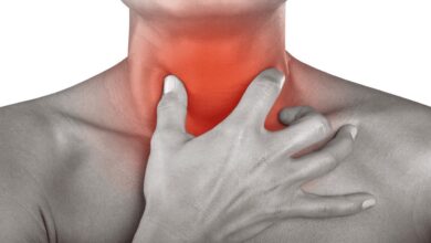
Angina pectoris (or angina)
Angina pectoris is characterized by oppressive pain that can be located in the chest, left arm and jaw.
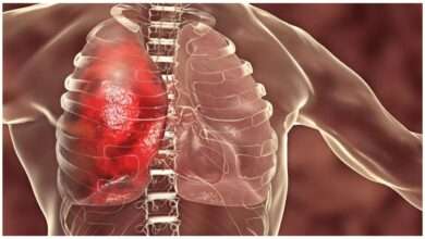
Myocardial infarction (or heart attack)
Infarction is the partial destruction of the heart muscle. The heart is then no longer able to play its role as a pump and stops beating.
Pericarditis
Pericarditis is an inflammation of the pericardium due to infections: viral, bacterial or parasitic. Inflammation can also occur after more or less significant trauma.
Valvulopathies
Valvulopathy is an alteration of the functioning of the valves of the heart by various diseases which can modify the cardiac function.
Heart rhythm disorders (or cardiac arrhythmia)
Heart rhythm disorders are characterized by irregular, too slow or too fast heartbeats, without these rhythm changes being linked to a so-called “physiological” cause (physical effort, for example).
Cardiomyopathies
Cardiomyopathies are diseases that cause dysfunction of the heart muscle, the myocardium. This disease causes a decrease in the muscle’s ability to pump blood and eject it into the circulation.
heart defects
Birth defects of the heart are present at birth.
Heart prevention and treatment
Risk factors
There are various factors that increase the risk of heart attack and stroke:
- smoking;
- a poor diet ;
- obesity;
- physical inactivity ;
- excessive alcohol consumption;
- hypertension;
- diabetes ;
- hyperlipidemia.
Prevention
The WHO recommends at least 30 minutes of physical activity per day.
Eating five fruits and vegetables a day and limiting salt intake also help prevent heart or stroke.
Anti-inflammatories (NSAIDs) and cardiovascular risks
Studies have shown that prolonged and high-dose intake of NSAIDs (Advil, Iboprfen, Voltaren, etc.) exposes people to cardiovascular risks.
Mediator and valve disease
Originally prescribed to treat hypertriglyceridemia (too high levels of certain fats in the blood) or hyperglycemia (too high sugar levels), the pick has also been prescribed for diabetics who are overweight.
The « appetite suppressant » property of the pick has led to it being widely consumed outside these indications to help non-diabetic people lose weight. It was then associated with valvulopathies and a rare cardiovascular pathology called Pulmonary Arterial Hypertension (PAH).
Heart tests and examinations
Your doctor will first carry out a basic examination: reading the blood pressure, listening to the beating of the heart, taking the pulse, evaluating the breathing or auscultating the abdomen.
Doppler ultrasound
Doppler ultrasound is a medical imaging technique that examines the conditions of flow and irrigation of the heart and blood vessels in order to check the obstruction of the arteries or the condition of the valves.
Coronographie
Coronagraphy is a medical imaging technique that visualizes the coronary arteries.
Ultrasound of the heart (or echocardiography)
Ultrasound of the heart is a medical imaging technique that allows the visualization of the internal structures of the heart (cavities and valves).
Electrocardiography at rest or under stress
Electrocardiography is a test that helps record the electrical activity of the heart to detect abnormalities.
Cardiac scan
Cardiac scintigraphy is an imaging test that observes the quality of the blood supply to the heart by the coronary arteries.
CT angiography
CT angiography is an examination that allows you to explore the blood vessels to detect a pulmonary embolism, for example.
Bypass surgery
Coronary bypass is a surgical procedure performed when the coronary arteries are blocked in order to restore circulation.
Lipid profile
- Dosage of triglycerides: in excessive quantity in the blood, they can contribute to the obstruction of the arteries;
- Cholesterol assay: LDL-cholesterol, referred to as “bad” cholesterol, is associated with an increased cardiovascular risk when it is present in excessive amounts in the blood;
- Fibrinogen assay : it is useful for monitoring the effect of a so-called “ fibrinolytic” treatment , intended to dissolve a blood clot in the event of thrombosis .
History and symbolism of the heart
The heart is the most symbolic organ of the human body.
During Antiquity, it was seen as the center of intelligence. Then, it was perceived in many cultures as the seat of emotions and feelings, perhaps because the heart reacts to an emotion and also provokes it.
It was in the Middle Ages that the symbolic shape of the heart appeared. Globally understood, it translates passion and love.