the heart
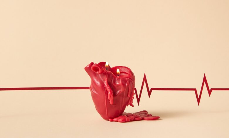
The heart (from the Greek word cardia and from the Latin cor, “heart”) is the central organ of the cardiovascular system. A real “pump”, it ensures the circulation of blood in the body thanks to its rhythmic contractions.
In close connection with the respiratory system, the heart allows the oxygenation of the blood and the elimination of carbon dioxide (CO2). Focus on the heart, its pathologies and possible treatments.
heart anatomy
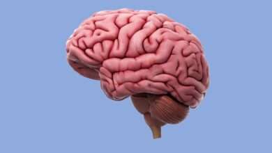
The heart is a hollow, muscular organ located in the rib cage.
Located between the two lungs at the back of the sternum, the heart is shaped like an inverted pyramid. Its vertex (or apex) rests on the diaphragm muscle and points downward, forward, to the left.
No bigger than a closed fist, the heart weighs on average 250 to 350 g in adults for about 12 cm in length.
Envelope and wall of the heart
The heart is surrounded by an envelope, the pericardium. It is made up of two layers: one is attached to the heart muscle, the myocardium, and the other stably attaches the heart to the lungs and the diaphragm.
The wall of the heart is made up of three layers, from outside to inside:
- the epicardium;
- the myocardium, it constitutes the bulk of the mass of the heart;
- the endocardium, which lines the cavities.
The irrigation of the heart is ensured on the surface by the system of coronary arteries which supply it with the oxygen and nutrients necessary for its proper functioning.
cavities of the heart
The heart is divided into four chambers: two atria (or atria) and two ventricles. Coupled two by two, they form the right heart and the left heart. The atria are located in the upper part of the heart, they are cavities for receiving venous blood.
In the lower part of the heart, the ventricles are the starting point for blood circulation. By contracting, the ventricles pump blood out of the heart into different vessels.
The cavities are the real pumps of the heart. Their wall is thicker than that of the atria and alone represent almost the entire mass of the heart.
The atria are separated by a partition called the inter-atrial septum and the ventricles by the inter-ventricular septum .
Heart valves
In the heart, four valves give blood a one-way flow.
Each atrium communicates with the corresponding ventricle via a valve: the tricuspid valve on the right and the mitral valve on the left.
The other two valves are located between the ventricles and the corresponding artery: aortic valve and pulmonary valve. Acting as a sort of “valves”, the valves prevent the backflow of blood as it passes between two cavities.
Physiology of the heart
Double pump
The heart, thanks to its role as a suction and repression double pump, ensures the circulation of blood in the body to bring oxygen and nutrients to the tissues.
There are two types of circulation: pulmonary circulation and systemic circulation.
Pulmonary circulation
The pulmonary circulation or small circulation has the function of transporting blood to the lungs in order to ensure gas exchange and then to bring it back to the heart. The right side of the heart is the pump of the pulmonary circulation.
Oxygen-depleted, CO2-rich blood enters the right atrium from the body through the inferior and superior vena cava.
The blood descends into the right ventricle which ejects it into the two pulmonary arteries (pulmonary trunk).
The pulmonary arteries then carry blood to the lungs where it gets rid of CO2 and absorbs oxygen. The oxygen is then redirected to the heart, in the left atrium, through the pulmonary veins.
Systemic circulation
Systemic circulation ensures the general distribution of blood to tissues throughout the body and its return to the heart. Here, it is the left heart that acts as a pump.
The reoxygenated blood arrives in the left atrium and then passes to the left ventricle, which ejects it by contraction into the aorta artery. From there, the blood is distributed to the various organs and tissues of the body. It is then brought back to the right heart by the venous network.
Heartbeat and spontaneous contraction
Circulation is provided by the beating of the heart.
Each beat corresponds to a contraction of the heart muscle, the myocardium, which is made up of large parts of muscle cells.
Like all muscles, the heart contracts under the influence of successive electrical impulses. But, the heart has the particularity of contracting spontaneously, rhythmically and independently thanks to an internal electrical activity.
The average heart beats 3 billion times over a lifetime of 75 years.