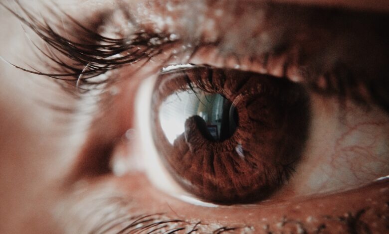
The eye (from the Latin word oculus, eye) is the organ of vision. By capturing light, it allows us to interact with the environment. The human eye can distinguish shapes and colors.
eye anatomy
The adult eye, or eyeball, is a sphere about 2.5 cm in diameter. It is protected in front by the eyelids, each of which bears eyelashes. They are lined by a membrane, the conjunctiva, which secretes a lubricating mucus to prevent the eye from drying out. The lacrimal gland, located above the eye, continuously releases tears (about 1mL/day/eye) helping to clean and protect the eye.
The bulb of the eye
The eye itself is called the bulb of the eye. It is a hollow sphere filled with liquids whose wall is made up of three layers (envelopes) superimposed. The transparent lens is the “lens” of the eye, it focuses light on the retina. It is protected by a liquid called aqueous humor.
The outer envelope of the eye is a fibrous tunic. It is a protective membrane which is composed of two elements: the sclera and the cornea. The sclera is the “white of the eye”. The cornea is transparent, it is the most exposed part of the eye. She very often suffers injuries but her ability to regenerate and heal is extraordinary. The cornea can also be transplanted without risk of rejection because it does not contain any blood vessels.
The intermediate envelope is a vascular tunic which is made up of three elements:
- The choroid, behind the eye, is a membrane rich in blood vessels which nourishes the three ocular tunics. It contains a dark brown pigment that absorbs light preventing its reflection inside the eye.
- The ciliary body is located in front of the eye, in continuity with the choroid. Composed of ciliary muscles, it acts on the shape of the lens by adapting its curvature to the distance from objects.
- The iris is located between the cornea and the lens. It contains pigment cells that determine the color of the eyes. In the center is the pupil, a round hole that lets light into the eye.
The inner coat of the eye is the retina. It extends from back to front, to the ciliary body. Half a millimeter thick, it is composed on its outer side of a pigmentary layer, glued to the choroid, which absorbs excess light thanks to melanin granules. Its inner side contains millions of visual cells: rods and cones. In its center, there is a small area called the macula : this is where visual acuity is maximum.
Visual cells and light in the eye
Visual cells
Visual cells are called photoreceptor neurons because they react to light.
The rods (120 million in each retina) are largely located at the periphery of the retina. They are responsible for peripheral vision and make it possible to distinguish shades of gray in the dark.
The cones (6 million) are mainly located in the center of the retina. They are involved in the vision of colors and details in bright light.
There are three types, each of which detects different wavelengths of visible light:
- The cones of the first type react mainly to blue light
- The second type cones in green light
- Third type cones react to red light
When excited by light, photoreceptors translate light energy into an electrical signal. These visual messages leave the retina via the optic nerve to reach the brain where they will be interpreted to produce visual sensations.
Light path in the eye
When light enters the eye, it successively passes through the cornea, the aqueous humor, the lens and the vitreous body, then reaches the retina in order to stimulate the photoreceptors. The image projected on the retina is inverted and smaller than the observed object.


